Myths about teaching can hold you back

- Year 10
- OCR
- Higher
Observing mitosis in plant cells using a light microscope
I can use a light microscope to observe plant cells in different stages of mitosis.
- Year 10
- OCR
- Higher
Observing mitosis in plant cells using a light microscope
I can use a light microscope to observe plant cells in different stages of mitosis.
Lesson details
Key learning points
- A light microscope can be used to observe plant cells in different stages of mitosis (e.g. from onion root tip).
- The parts of a light microscope and their functions.
- The sequence of steps for setting up a light microscope to observe cells, including changing the magnification & focus.
- Explaining observations from microscopy using ideas about what happens during stages of the cell cycle.
Keywords
Light microscope - A type of microscope that uses visible light and a system of lenses to generate magnified images of small objects.
Mitosis - A type of cell division that produces genetically identical cells.
Lens - A piece of glass or other transparent material with curved sides, used in a microscope to magnify objects.
Magnification - Making small objects appear larger in order to see more detail.
Focus - A point where light rays converge to form an image with clarity.
Common misconception
Recognising that after cell growth DNA condenses to form visible chromosomes and the nuclear membrane breaks down.
There are slides that aim to show the difference between DNA held within the nucleus and visible chromosomes where DNA has condensed. There is also a check for understanding question and practice task question to reinforce this concept.
To help you plan your year 10 biology lesson on: Observing mitosis in plant cells using a light microscope, download all teaching resources for free and adapt to suit your pupils' needs...
To help you plan your year 10 biology lesson on: Observing mitosis in plant cells using a light microscope, download all teaching resources for free and adapt to suit your pupils' needs.
The starter quiz will activate and check your pupils' prior knowledge, with versions available both with and without answers in PDF format.
We use learning cycles to break down learning into key concepts or ideas linked to the learning outcome. Each learning cycle features explanations with checks for understanding and practice tasks with feedback. All of this is found in our slide decks, ready for you to download and edit. The practice tasks are also available as printable worksheets and some lessons have additional materials with extra material you might need for teaching the lesson.
The assessment exit quiz will test your pupils' understanding of the key learning points.
Our video is a tool for planning, showing how other teachers might teach the lesson, offering helpful tips, modelled explanations and inspiration for your own delivery in the classroom. Plus, you can set it as homework or revision for pupils and keep their learning on track by sharing an online pupil version of this lesson.
Explore more key stage 4 biology lessons from the Cell division: mitosis and meiosis unit, dive into the full secondary biology curriculum, or learn more about lesson planning.

Equipment
Light microscopes, prepared slides.
Licence
Lesson video
Loading...
Prior knowledge starter quiz
6 Questions
Q1.What word describes making objects appear larger using a lens?
Q2.What are the smallest living units of life?
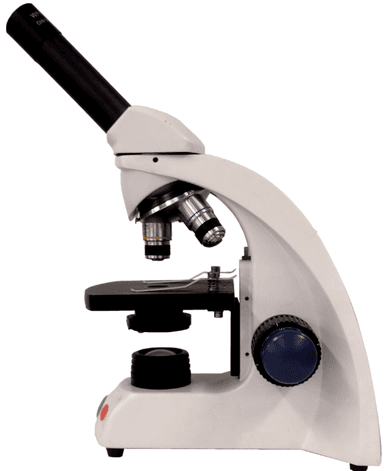
Q3.What is this piece of scientific equipment called?
Q4.What is mitosis?
Q5.Match each stage of the cell cycle to the description of what happens?
DNA and sub-cellular structures are copied.
Chromosomes line up in the middle, are pulled apart. Nucleus divides.
Division of cytoplasm and cell membrane.
Q6.Which of the following is not true for mitosis?
Assessment exit quiz
6 Questions
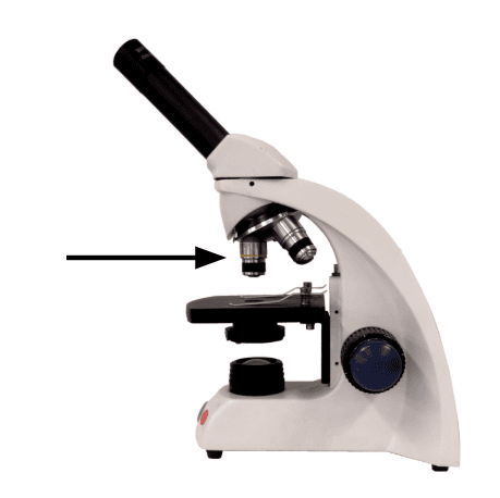
Q1.Which part of the microscope is the line pointing towards?
Q2.In which two places does mitosis take place in an onion?
Q3.Put these stages in order to show how a microscope should be used?
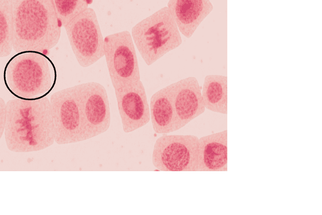
Q4.The image has circled a stage of the cell cycle, what is happening?
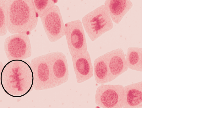
Q5.The image has circled a stage of the cell cycle, what is happening?