Myths about teaching can hold you back
- Year 11
- OCR
- Higher
Light microscopy: observing and identifying microorganisms
I can explain how microorganisms can be observed and identified using a light microscope.
- Year 11
- OCR
- Higher
Light microscopy: observing and identifying microorganisms
I can explain how microorganisms can be observed and identified using a light microscope.
These resources were made for remote use during the pandemic, not classroom teaching.
Switch to our new teaching resources now - designed by teachers and leading subject experts, and tested in classrooms.
Lesson details
Key learning points
- A light microscope can be used to observe microorganisms.
- Observations from a light microscope can be used to identify microorganisms, including pathogens causing a disease.
- The observed shape of a microorganism can help to identify it.
- Use of a key and stains can help to identify a microorganism.
- Growing microorganisms in specific conditions can also help to identify them.
Keywords
Microorganism - A living thing that is too small to see with the unaided eye.
Light microscope - A type of microscope that uses visible light and a system of lenses to generate magnified images of small objects.
Pathogen - A microorganism that causes disease.
Key - A series of questions about the features of an organism that help us to identify it.
Stain - A coloured liquid put onto a specimen so that the cells and their structures can be more easily seen with a light microscope.
Common misconception
Students often use the term "zoom in" instead of focus.
You must correct the term "zoom in" to focus. You are not making the image larger, you are focussing to make the image clearer.
To help you plan your year 11 biology lesson on: Light microscopy: observing and identifying microorganisms, download all teaching resources for free and adapt to suit your pupils' needs...
To help you plan your year 11 biology lesson on: Light microscopy: observing and identifying microorganisms, download all teaching resources for free and adapt to suit your pupils' needs.
The starter quiz will activate and check your pupils' prior knowledge, with versions available both with and without answers in PDF format.
We use learning cycles to break down learning into key concepts or ideas linked to the learning outcome. Each learning cycle features explanations with checks for understanding and practice tasks with feedback. All of this is found in our slide decks, ready for you to download and edit. The practice tasks are also available as printable worksheets and some lessons have additional materials with extra material you might need for teaching the lesson.
The assessment exit quiz will test your pupils' understanding of the key learning points.
Our video is a tool for planning, showing how other teachers might teach the lesson, offering helpful tips, modelled explanations and inspiration for your own delivery in the classroom. Plus, you can set it as homework or revision for pupils and keep their learning on track by sharing an online pupil version of this lesson.
Explore more key stage 4 biology lessons from the Defences against pathogens, the human immune system and vaccination unit, dive into the full secondary biology curriculum, or learn more about lesson planning.

Content guidance
- Risk assessment required - equipment
Supervision
Adult supervision required
Licence
Prior knowledge starter quiz
6 Questions
Q1.What do we call a series of questions used to help identify organisms?

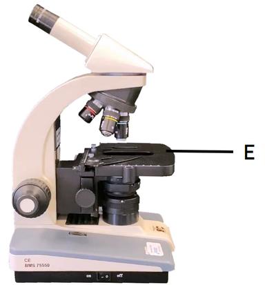
Q2.Which part of the microscope is labelled E?
Q3.True or false? Bacteria are multicellular prokaryotes.
Q4.Which type of drug is a doctor likely to prescribe to a patient with a bacterial infection?
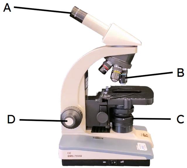
Q5.Which part of the microscope would you use to bring your specimen into focus?
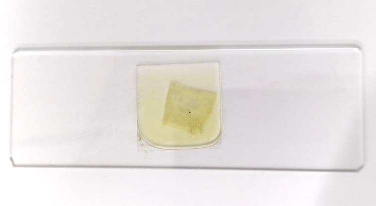
Q6.Put these steps in order to explain how to make a temporary mount of onion tissue.
Assessment exit quiz
6 Questions
Q1.Match the part of the microscope to its description.
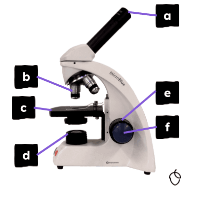
Eyepiece lens: viewing lens with ×10 magnification.
Objective lenses: three lenses with different magnifications.
Stage: specimen or slide is placed here.
Light source: illuminates the specimen so that it can be observed.
Coarse focus wheel: for adjusting the focus in larger increments.
Fine focus wheel: for adjusting the focus in smaller increments.
Q2.Put these steps in order to explain how a light microscope is set up.

Q3.How can you increase the magnification of a light microscope?
Q4.What can we use to make living material easier to see under a microscope?
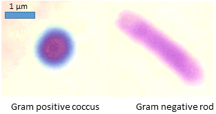
Q5.We can use a to identify types of bacteria.

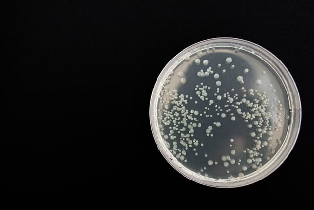
Q6.The image below shows bacteria growing on an agar plate containing penicillin. Penicillin works best on gram-positive bacteria. What can we conclude about the bacteria growing on the plate?
